Lab 10 - Leaves I
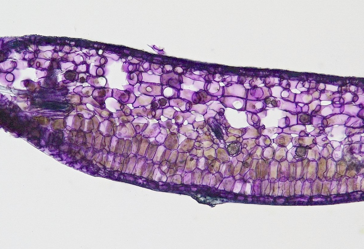
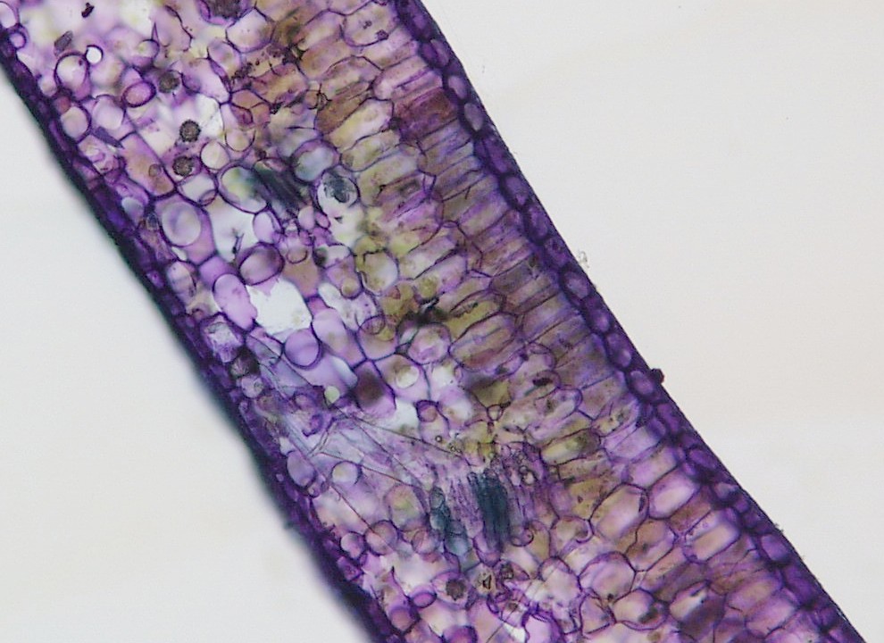
Here are a few images made from sections obtained by students in the
first leaves lab. The first six are English ivy leaf
sections. You can see the palisade layers and large air spaces in
the spongy mesophyll fairly well. Vascular tissue is not clearly
visible, but you should be able to figure out adaxial and abaxial from
the mesophyll layers.



Next are four images of Mulberry leaves.

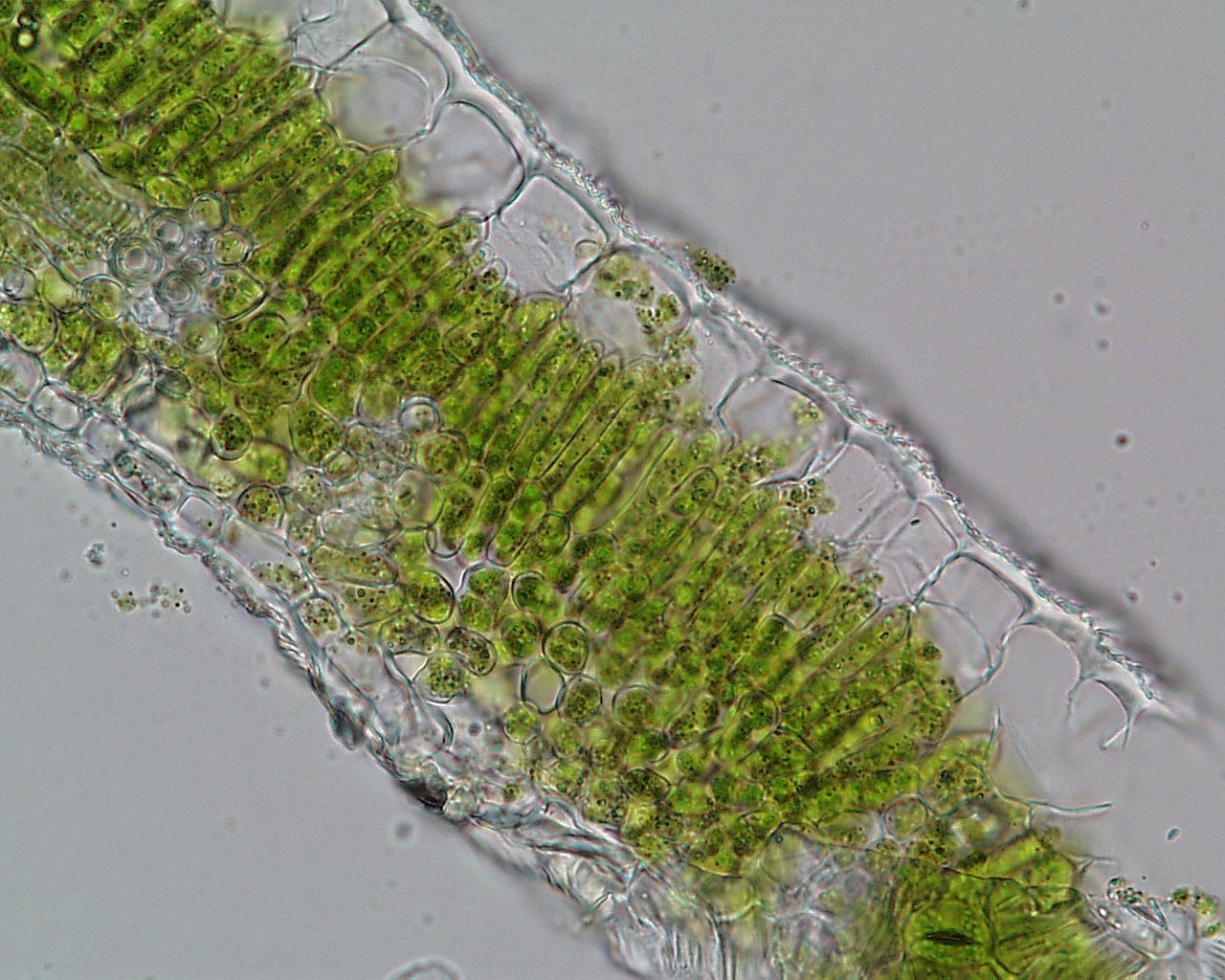

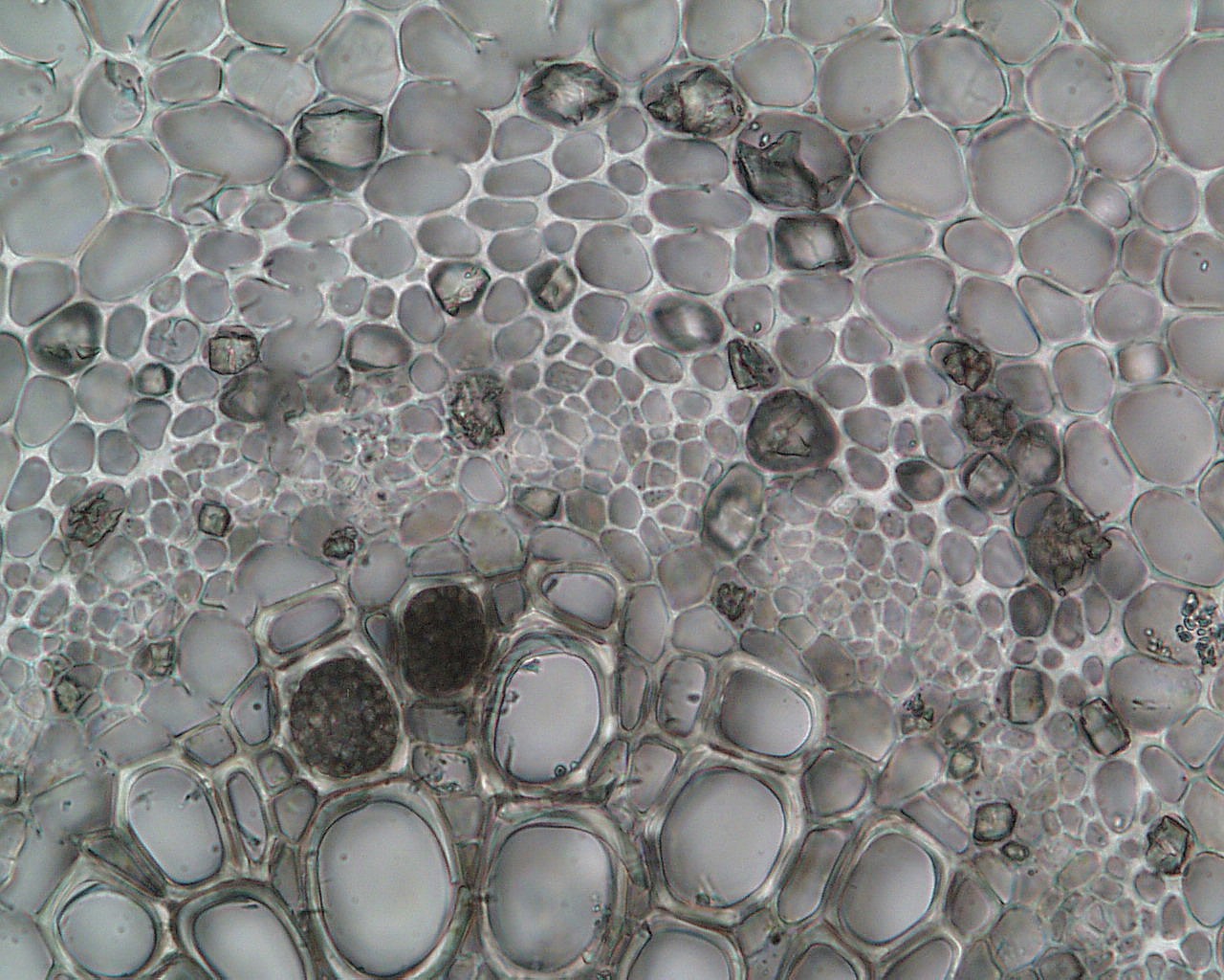
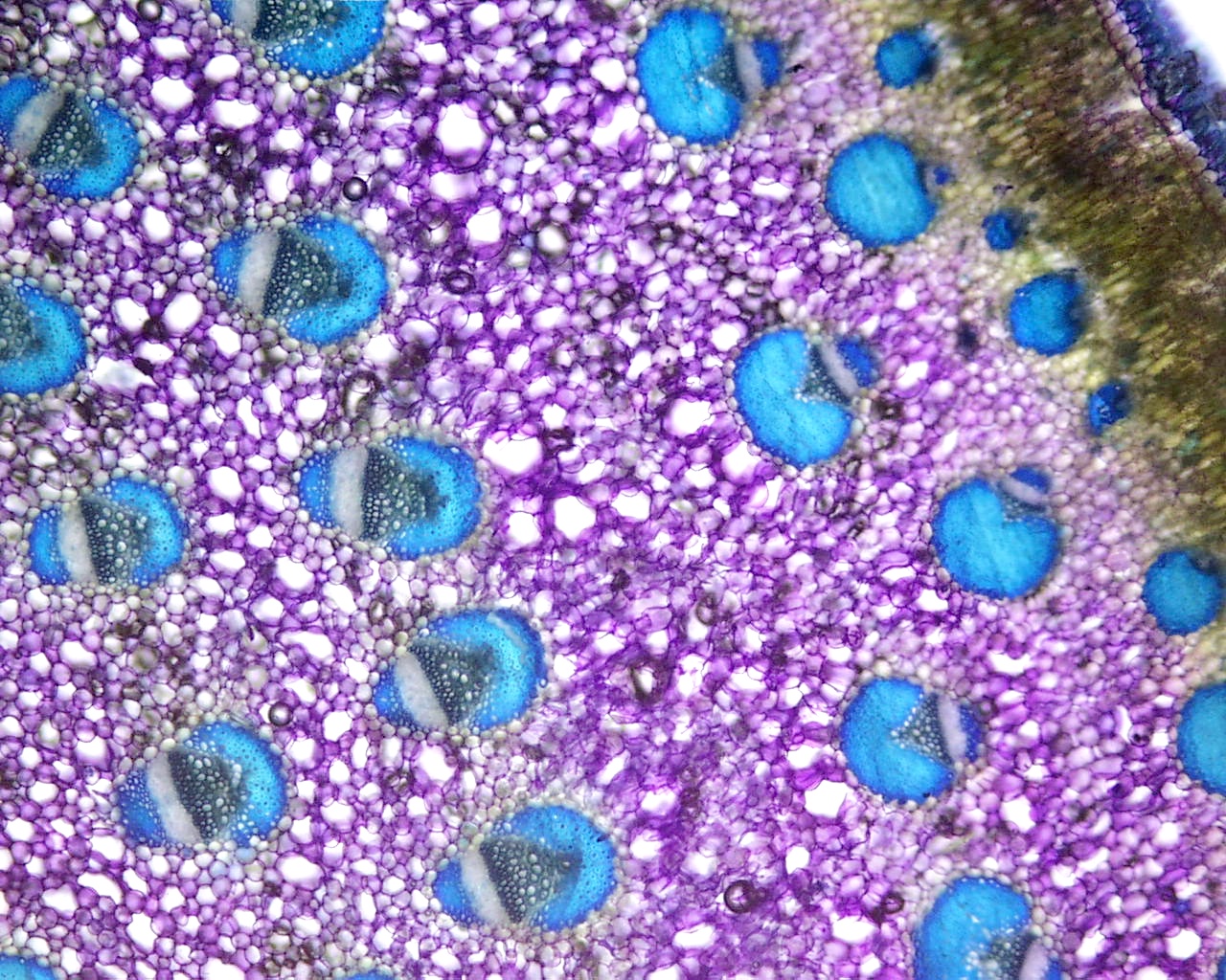
Here is a wide view of the center of a yucca leaf. Notice the
vascular bundle orientations - this is a unifacial leaf!
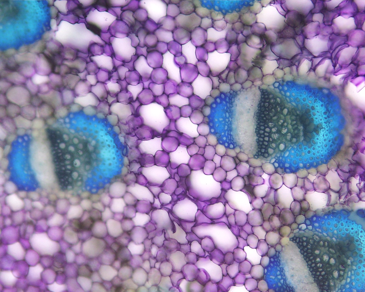
Here's a closer view that might help you pick out the xylem, phloem,
and fibers in each bundle. Nice clean section!
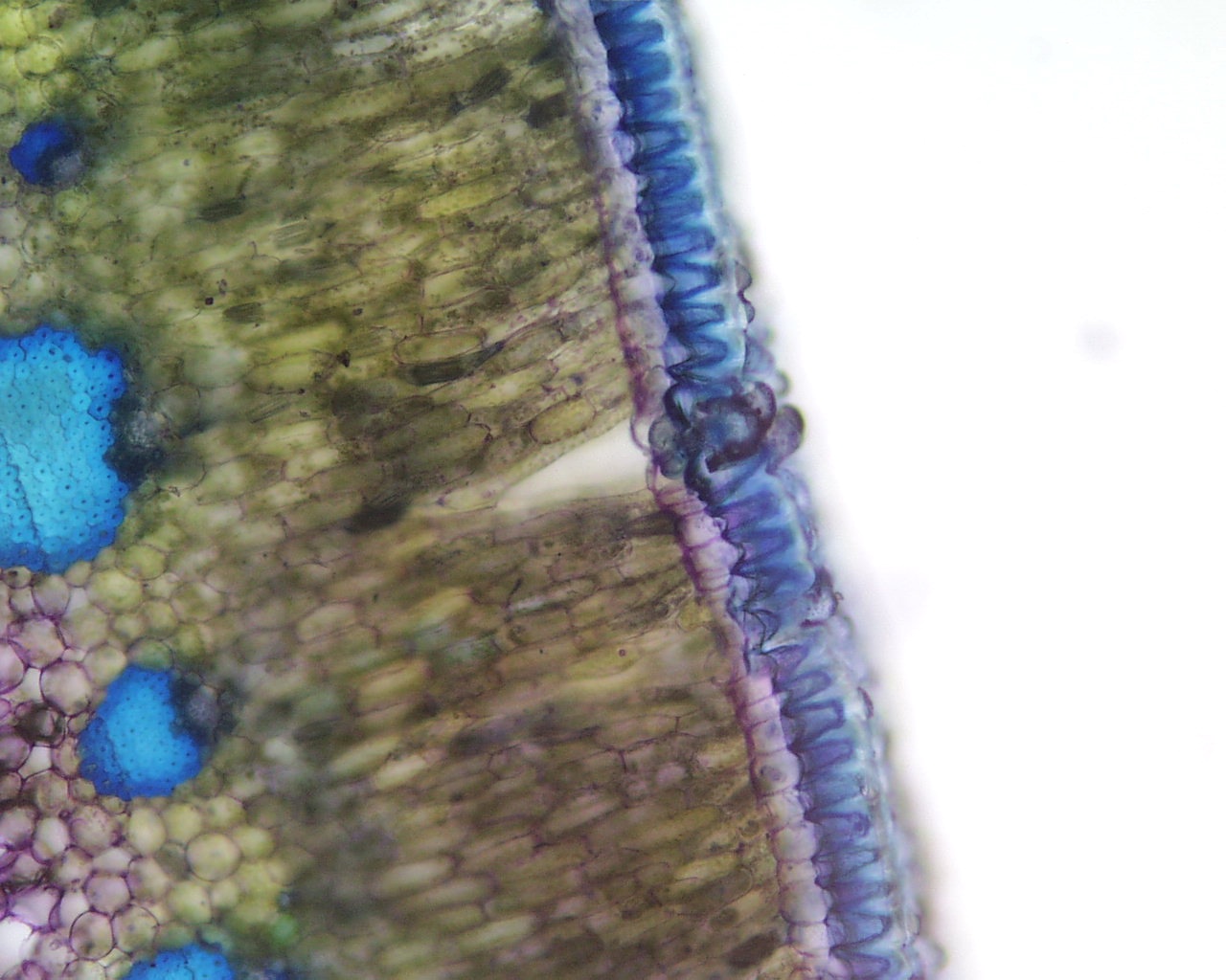
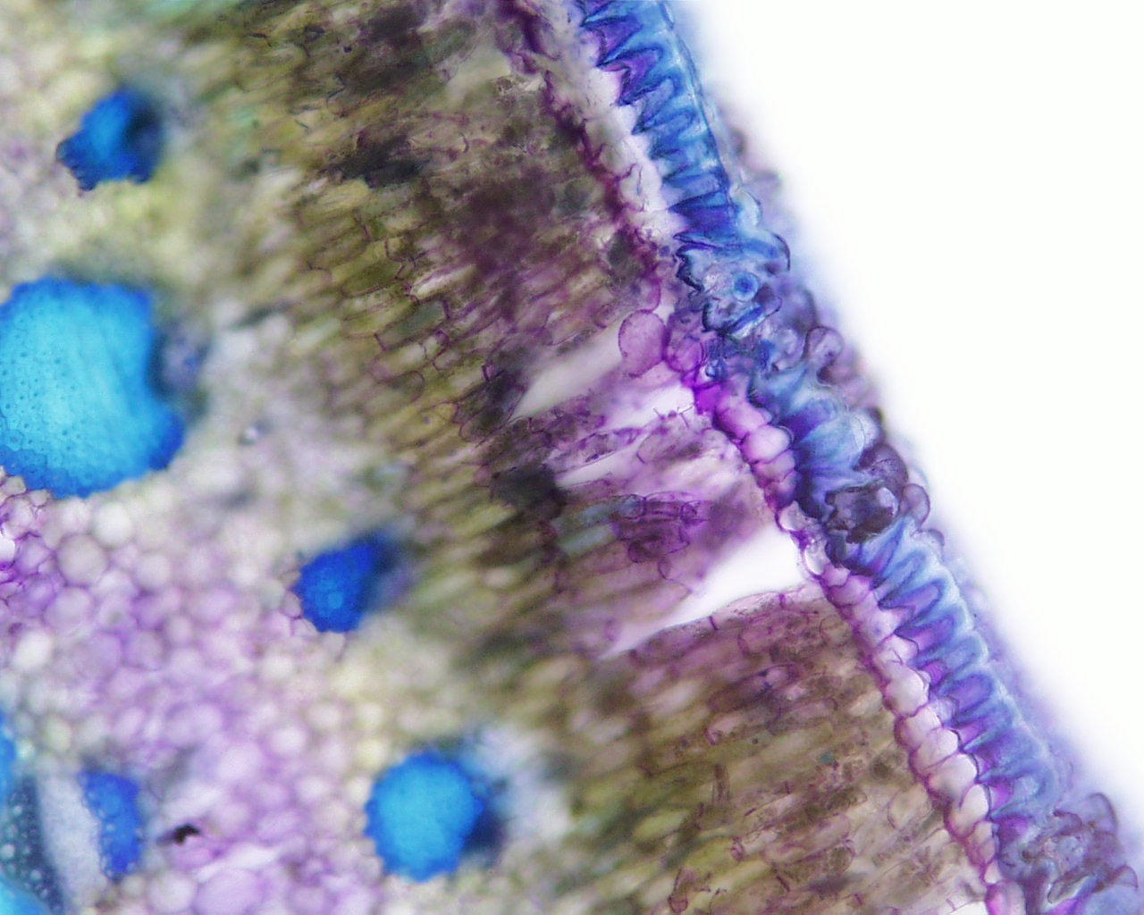
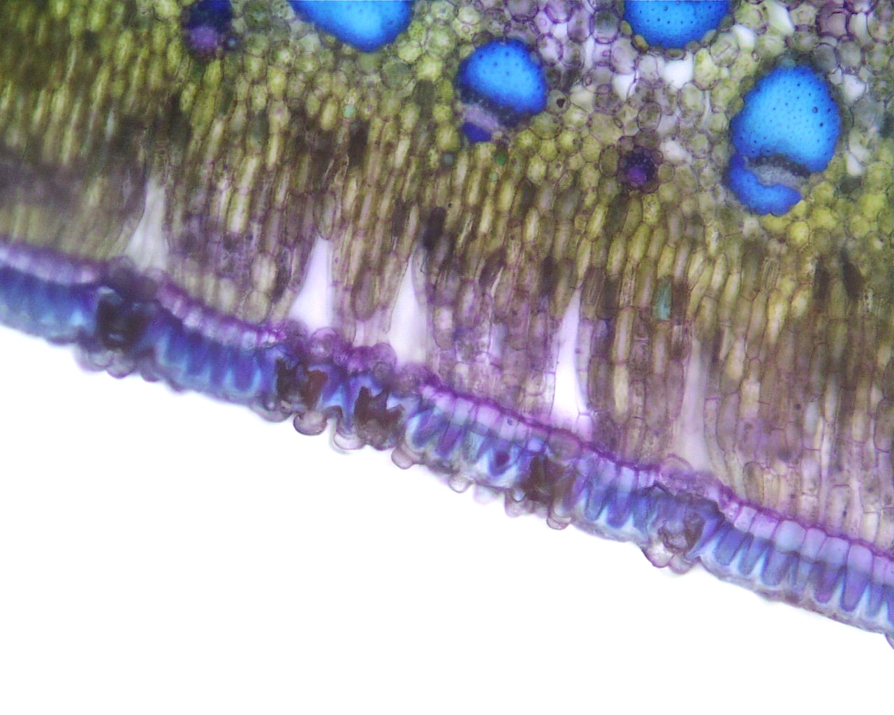
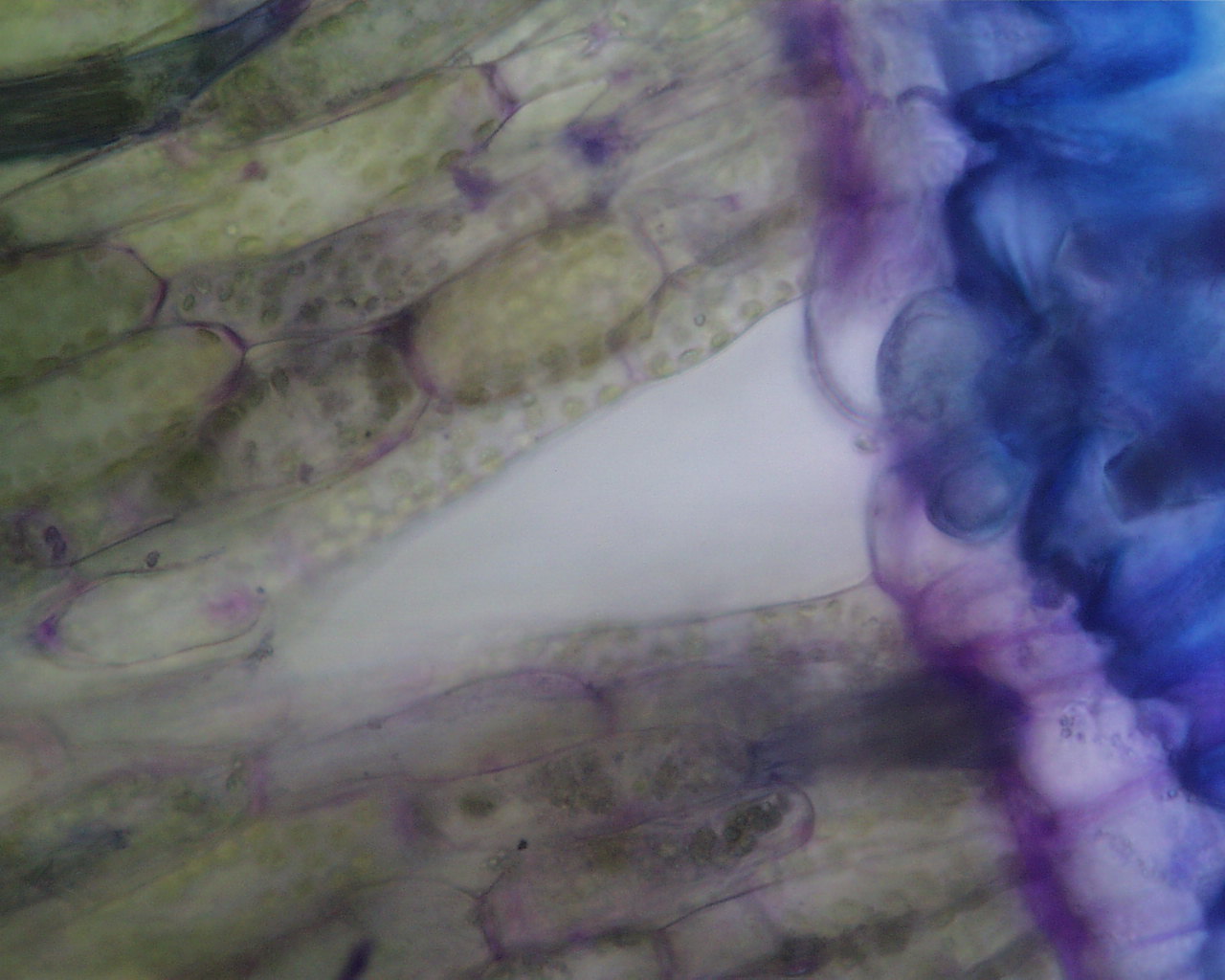
These last four images show the outer regions of the yucca leaf.
You can see the palisade layers, the epidermis, and stomata with their
sub-stomatal cavities very nicely. Especially in the last image
below, you can really see those chloroplasts nicely!